Describe the Uses of Dental Imaging
Advantages and disadvantages of various imaging techniques in endodontic are presented. Today there are various types of x-rays and other imaging methods available that can be used to detect.

Coconut Oil For Dental Health And Neurodegenerative Disorders Dental Health Dental Coconut Oil
Ectopic teeth periapical radiographs can be used to identify and localise ectopic teeth using a parallax technique where 2 periapicals are taken in different positions to assess the location.
. A dental 3D scan allows clinicians to view dental anatomy from different angles. The uses of dental imaging are checking patients oral health and making it clearer to diagnose certain teeth problems such as cavities. Digital radiography is a type of X-ray imaging that uses digital X-ray sensors to replace traditional photographic X-ray film producing enhanced computer images of teeth gums and other oral structures and.
The history of dental imaging began in the late 1800s with the development of the x-ray image. One one-thousand 11000 of an ampere a unit of measurement used to describe the instensity of an electrical current. During the last three decades this technology has improved tenfold alongside the benefits that these systems provide.
The most commonly used in dentistry is the cbct or cone-beam technology. Dentists are using dental imaging to perform dental services that were unimaginable a few years ago. 3D Imaging Using CBCT.
Dental imaging helps our dentist make a thorough well-informed diagnosis and treatment plan catered to your unique needs. The application of computer imaging software used in dentistry has become one of the main tools for dentists in providing superior services to their. Periapical lesions which show that a tooth has infection can usually be.
Federal and state regulations. Dental X-ray tube acts as a self-rectifier in that it changes AC to DC while producing x-rays this ensures that the current is always flowing in the same direction from cathode to anode What circuits are involves with x-rays. Dental CBCT systems have been sold in the United States since the early 2000s and are increasingly used by radiologists and dental professionals for various clinical applications including.
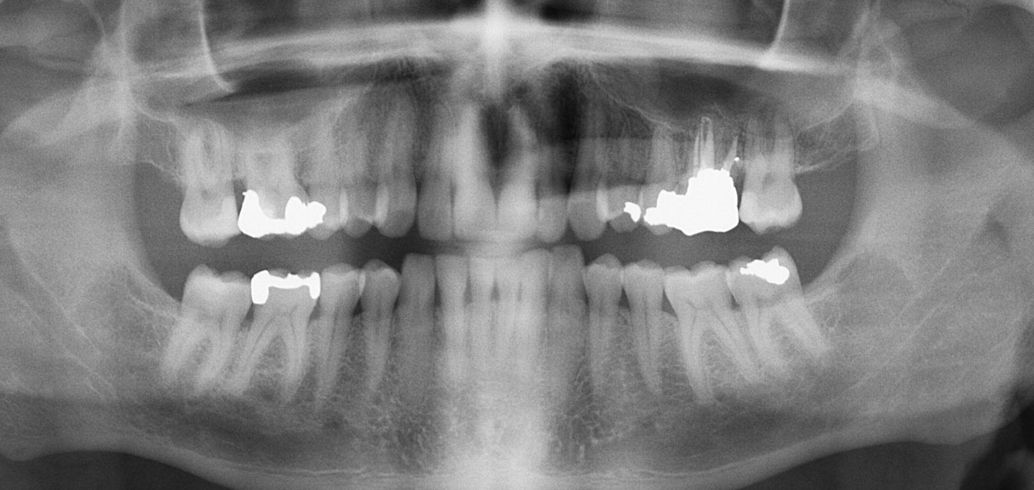
Most states have laws that require inspections of dental x-ray equipment on a regular basis such as every _____years. Dental x-rays help your dentist identify diseases and problems. Assessment of root morphology this may be important prior to dental extractions particularly for wisdom teeth.
The most common use of a cbct is to plan placement of dental implants. The improvements in imaging technology have helped in obtaining a near perfect image for accurate diagnosis. Digital imaging Dental Film and Processing Radiographs flashcards.
Only through the use of dental radiographs. Orthodontists and dentists use Cone Beam Computerized Tomography CBCT which evolved from CAT. Describe the discovery of x-radiation.
3D dental imaging is rapidly becoming the standard of care in all of dentistry not just specialty fields. Provide information during dental procedures such as root canal therapy Document a patients condition at a specific time. To find out how you can benefit from 3D images make an appointment by calling our office at 951 244-9495 or going online today.
Hauser uses 3D scans to diagnose a wide variety of dental problems. In 1973 computed tomography CT created images by combining x-ray and computer technology to capture thin slices of tissue1 After that magnetic resonance imaging MRI allowed soft tissue analysis. Also many dental offices are now using digital images instead of film.
Create flashcards for FREE and quiz yourself with an interactive flipper. Radiation health codes may include regulations pertaining to. Barriers film speeds position of the operator and film processing.
Many dental diseases and conditions have no clinical signs or sympstoms and are typically discovered. Additionally the scans are used in conjunction with computer aided design CAD programs and CEREC to craft precise dental restorations. Imaging plays an important role in endodontics and is routinely utilized for the following diagnosis.
If you are a doctor who looks at the precision of this equipment and thinks its overkill for the patients you see it may be time to rethink that assumption. Dental x-rays can also help your dentist evaluate injuries to your face and mouth. These scans are used to evaluate structures in three dimensions which is an advantage over traditional dental x-rays that are only two dimensional.
Radiographs enable the dentist to see. Periapical and cephalometric radiographs have. Dental radiographs help dentists assess the development of teeth and bones in children.
Identify bone loss in the early stages. Therefore imaging of these structures is one of useful diagnostic tools for clinicians to make decision treatment modality. Evaluate growth and development.
Conditions that are not visible in the oral cavity and to identify many conditions that might otherwise remain undetected. Detect dental caries in the early stages. A recording medium for an image normally film phosphor storage plate psp or a digital sensor.
Dental professionals today are increasingly using digital dental radiographs digital X-rays to better detect diagnose treat and monitor oral conditions and diseases. In the case of 3D dental imaging the advantages are clear granting practitioners and patients alike a better clinical experience. Locate abnormalities in surrounding hard and soft tissues.
Uses of digital imaging-To detect lesions diseases and conditions of teeth and surrounding structures-Confirm or classify suspected disease-Provide information during dental procedures root canal therapy instrumentation and surgical placement of. Describe the use of dental imaging. A 3D scan can help gain a better view of bone structures such as adjacent root positions in order to locate canals and root fractures as well as provide the ability to more.
Process by which electrons are removed from atoms causing harmful effects of radiation in humans. Not only is time saved in the development process reducing the amount of radiation by as much as 80. An offshoot of dental X-rays is 3D imaging.
The use of dental x-ray equpiment is regulated by. 3D imaging for orthodontic purposes contain pre- and post-treatment evaluation of dentoskeletal and craniofacial relationships and facial appearance and beauty inspecting treatment results in terms of soft and underlying hard tissues and 3D.

Types Of Dental Radiographs And Their Uses Dentalnotebook
What Is Opg X Ray Information About Uses Diagnosis And Treatment

Types Of Dental Radiographs And Their Uses Dentalnotebook


Comments
Post a Comment